

Know What
To Look For
O
Different, unusual, or just doesn't look right
D
Rough, flaky, crusted, or scab-like surface
D
Black, brown, pink, white, or combinations
S
Translucent with tiny blood vessels visible
P
Raised, swollen, tender, or painful to touch
O
Wound that bleeds, oozes, or won't close
T
New, growing, or changing in any way
More Than Just Melanoma
For decades, the ABCDE criteria focused public awareness almost entirely on melanoma — leaving basal cell and squamous cell carcinomas largely outside the patient education conversation. Yet BCC and SCC together make up roughly 95% of all skin cancers diagnosed each year. ODD SPOT™ was developed to close that gap: a single, memorable tool that helps patients recognize the warning signs of all three major skin cancers, backed by peer-reviewed science and designed for real-world use.
Know Your Cancers
MOST COMMON
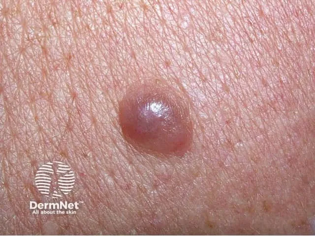
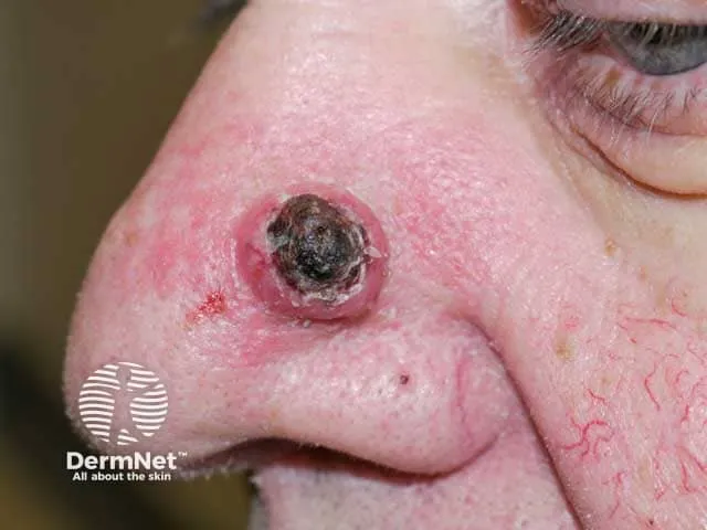
Basal Cell Carcinoma
3.6M
U.S. CASES/YR
~80%
OF SKIN CANCERS
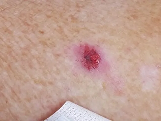
Basal cell carcinoma is the most common cancer in the United States, arising from the deepest layer of the outer epidermis. It grows slowly and rarely spreads to other organs — but untreated BCC can invade surrounding tissues, including nerves and bone.
BCC most often appears on sun-exposed areas such as the face, scalp, neck, and hands. It can present as a pearly or translucent bump, a flat scar-like lesion, a pink growth with raised edges, or a recurring open sore. Chronic UV exposure is the primary risk factor.
TREATMENT
Mohs surgery
Excision
ED&C
Topical therapy
Radiation
Targeted therapy
SECOND MOST COMMON
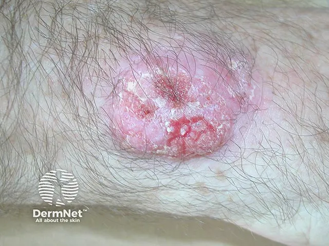
Squamous Cell Carcinoma
1.8M
U.S. CASES/YR
~80%
OF SKIN CANCERS
Squamous cell carcinoma is the second most common skin cancer, originating in the squamous cells that comprise the middle and outer layers of the skin. Unlike BCC, SCC carries meaningful metastatic potential — it can spread to lymph nodes or distant organs, particularly from high-risk sites such as the lip or ear.
SCC often develops from precancerous actinic keratoses caused by cumulative sun damage. It can appear as a firm red nodule, a flat keratotic plaque, a sore in an existing scar, or a wart-like growth. Immunosuppression, HPV, and chronic wounds are additional risk factors.
TREATMENT
Mohs surgery
Excision
Radiation
Cryotherapy
Immunotherapy
Chemotherapy
MOST DANGEROUS
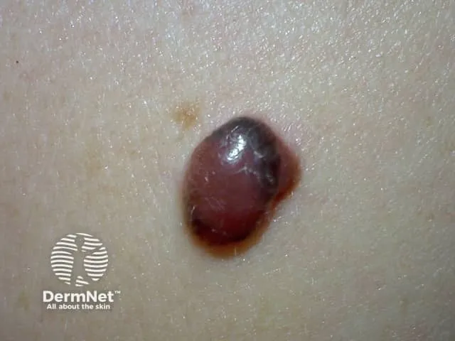
Melanoma
100K
U.S. CASES/YR
~1%
HIGHEST MORTALITY
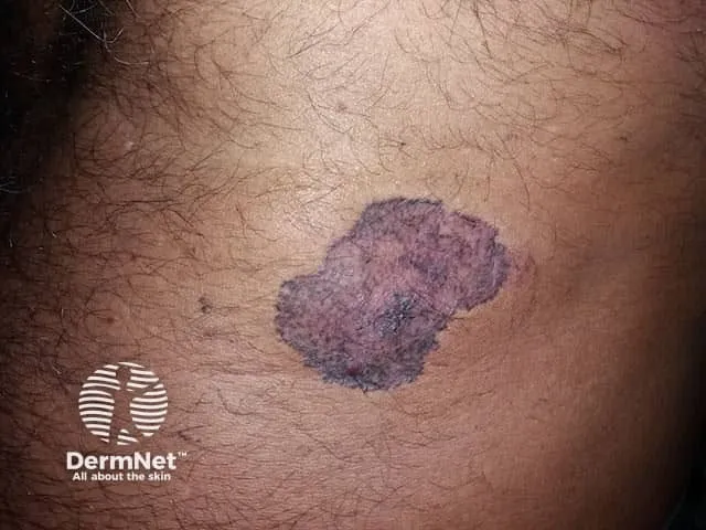
Melanoma is the most serious form of skin cancer, responsible for the majority of skin cancer deaths despite representing only about 1% of cases. It originates in melanocytes and can spread rapidly to lymph nodes, lungs, brain, and other organs when not caught early.
Melanoma can develop within an existing mole or appear as a new, unusual spot anywhere on the body — including areas never exposed to the sun. Risk factors include UV exposure, fair skin, family history, and a personal history of blistering sunburns. Survival rates are excellent with early-stage detection.
TREATMENT
Wide local excision
Sentinel node biopsy
Immunotherapy
Targeted therapy
Radiation
Clinical trials
ODD SPOT™ In Action
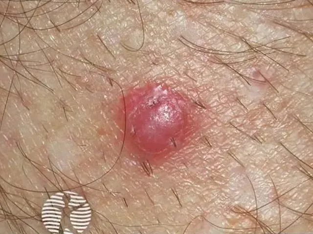
BASAL CELL CARCINOMA
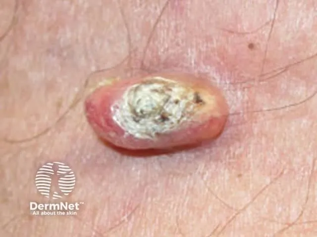
SQUAMOUS CELL CARCINOMA
MELANOMA
O
ODD LOOKING
BASAL CELL
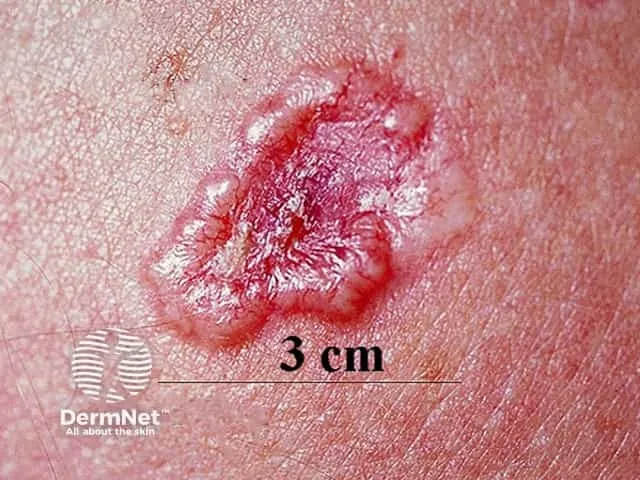
SQUAMOUS CELL
D
DRY / SCABBY
SQUAMOUS CELL
D
DISCOLORED
S
SHINY
BASAL CELL
SQUAMOUS CELL
MELANOMA
P
PUFFY / PAINFUL
BASAL CELL
SQUAMOUS CELL
MELANOMA
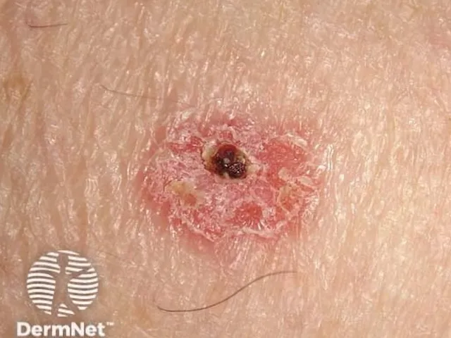
O
OPEN, OOZING OR BLEEDING
BASAL CELL
SQUAMOUS CELL
MELANOMA
T
TRANSFORMING
BASAL CELL
SQUAMOUS CELL